Otoplasty, commonly known as ear reshaping or prominent ear surgery, is a specialized facial plastic procedure designed to correct structural irregularities in the external ear framework. This surgery primarily addresses an underdeveloped outer ear fold (antihelical fold) or an excessively deep central ear bowl (conchal hypertrophy), both of which can cause the ears to protrude noticeably from the sides of the head. Undergoing otoplasty in Istanbul, Turkey, with an experienced facial plastic surgeon provides a permanent and well-proportioned adjustment. By making precise changes through an incision hidden entirely in the shadow fold behind the ear, this procedure restores a natural cranial angle, corrects structural asymmetries, and avoids an artificial, overly flattened appearance.
1. Introduction to Otoplasty (Ear Reshaping)
What is Otoplasty? Restoring Balance to the Craniofacial Framework
Otoplasty is a precise contouring surgery that refines the shape, position, and proportions of the external ear (auricle). Because the ears frame the face, significant asymmetry or protrusion can disrupt overall facial balance. This procedure goes beneath the surface skin to gently reshape the underlying cartilage framework, re-anchoring loose folds and reducing excessive depth to bring the ears into a balanced alignment with the head and facial features.
- Proportion Correction: Adjusts the angle of protruding ears to bring them into a natural, balanced position relative to the side of the head.
- Structural Reshaping: Rebuilds missing or flat ear folds, giving the cartilage a smooth and natural curvature.
- Hidden Incision Access: Incisions are placed completely within the natural crease behind the ear, ensuring no visible marks remain from the front or sides.
Anatomical Causes of Prominent Ears: Antihelical Underdevelopment and Conchal Hypertrophy
Prominent ears are typically caused by two main anatomical variations. The first is an underdeveloped antihelical fold, where the outer cartilage rim fails to fold back properly, causing the top portion of the ear to stick out. The second is conchal hypertrophy, an overgrowth or excessive depth of the central cartilage bowl that pushes the entire ear away from the skull. A thorough physical assessment allows the surgeon to identify these specific structural causes and customize the technique accordingly.
2. The Technical Science of Post-Auricular Incision & Cartilage Remodeling
Reshaping ear cartilage requires a careful blend of gentle structural modification and secure internal stitching to ensure the ear permanently holds its new position.
Concealing the Pathway Within the Post-Auricular Crease
The surgery begins with a precise incision placed entirely within the post-auricular crease—the natural groove where the back of the ear meets the head. This strategic entry path gives the surgeon direct access to the cartilage matrix while ensuring the resulting scar stays completely hidden within the natural shadows of the head.
The Mustarde Technique: Creating the Antihelical Fold
To rebuild a flat or missing outer ear fold, surgeons utilize the specialized Mustarde technique. This approach involves placing a series of permanent, non-absorbable mattress sutures through the back of the cartilage. As these sutures are carefully tightened, they guide the flat cartilage into a smooth, natural-looking antihelical curve, restoring a balanced shape without cutting completely through the ear structure.
Technical Comparison Matrix: Cartilage Scoring vs. Suture-Only Sparing Techniques
| Procedural Parameter | Traditional Cartilage Scoring (Incision-Heavy) | Suture-Only Cartilage Sparing (Advanced Approach) |
|---|---|---|
| Primary Mechanism | Cuts or thins the cartilage surface to break its structural memory. | Uses precise permanent internal mattress stitches to reshape the fold. |
| Cartilage Vitality | Higher risk of surface roughness or micro-irregularities. | Preserves the intact cartilage structure, keeping the surface completely smooth. |
| Contour Naturalness | Can sometimes create sharp, rigid, or artificial-looking edges. | Recreates soft, smooth curves that mimic natural ear anatomy perfectly. |
| Recovery Profile | Prone to increased swelling, internal bruising, and longer healing times. | Offers a gentler recovery with less localized swelling and faster tissue bonding. |
 Before & after — Otoplasty (Ear Reshaping)
3. Structural Scaling: Managing Conchal Hypertrophy & Mastoid Anchoring
When the central bowl of the ear is excessively deep, reshaping the outer fold alone is not enough. The surgeon must reduce this deeper volume to sit the ear closer to the head naturally.
Conchal Reduction: Trimming Excessive Cartilage Depth
In cases of pronounced conchal hypertrophy, a precise crescent-shaped piece of excess cartilage is removed from the deep bowl through the hidden incision behind the ear. This step directly lowers the height of the inner bowl walls, allowing the entire middle section of the ear to drop back closer to the side of the head without altering the appearance of the ear canal opening.
Mastoid Periosteal Anchoring: Preventing Recurrence
To ensure the ear stays securely in its new position, the surgeon uses strong, permanent sutures to anchor the conchal cartilage directly to the mastoid periosteum—the tough, fibrous bone lining behind the ear. This deep anchoring system counteracts the natural spring-like memory of the cartilage, providing a stable support system that prevents the ear from tilting forward again over time.
- No “Pinned-Back” Look: The positioning is calculated to leave a natural space (typically 1.5 to 2 centimeters) between the outer ear rim and the scalp.
- Symmetry Calibration: Measurements are verified on both sides during surgery to correct existing differences and ensure balanced facial symmetry.
4. Preventing Complications: Protecting Cartilage Vitality & Avoiding Hematoma
Ear cartilage is a delicate tissue that relies completely on its thin overlying skin and blood vessel network for nutrition, requiring precise handling during surgery.
The Critical Importance of Hemostasis
Because the skin over the ear cartilage is thin and highly vascular, careful fluid and bleeding control (hemostasis) is essential. If blood collects beneath the skin layers after surgery, it can form a hematoma. Left untreated, a hematoma can disrupt the blood supply to the cartilage. Surgeons prevent this by sealing blood vessels instantly during dissection and applying a specialized compression dressing immediately after closure.
Preserving Perichondrial Vascular Networks
The perichondrium is the thin membrane covering the cartilage that carries its vital blood supply. The surgical dissection is performed precisely within a safe tissue plane to keep this membrane intact, protecting the cartilage vitality, ensuring healthy tissue bonding, and minimizing the risk of long-term structural warping.
Age Groups vs. Cartilage Elasticity & Surgical Approach
| Patient Age Group | Cartilage Material Characteristics | Surgical Strategy Focus |
|---|---|---|
| Pediatric Patients (Ages 5 - 12) | Highly soft, elastic, and pliable cartilage matrix with minimal resistance. | Reshapes easily using gentle suture-only folding techniques; cartilage responds quickly with exceptional long-term stability. |
| Adult Patients (Ages 13 and Above) | Firmed, thicker, and more rigid cartilage framework with strong structural memory. | Requires a combination of internal conchal reduction, deep mastoid anchoring, and meticulous suture tension to secure the reset. |
5. The Surgical Journey with Prof. Dr. Gürkan Kayabaşoğlu
Reshaping the external ear structure requires precise calibration of facial angles and careful tension management. In our Istanbul clinic, each otoplasty is planned to match the patient’s individual cranial proportions, ensuring a natural result that maintains a healthy, soft curvature from every viewing angle.
The Otoplasty Clinical Workflow Steps
- Pre-Operative Geometric Measurement: The surgeon measures the auriculocephalic angle and the distance between the outer ear rim and the scalp, mapping out the precise structural changes needed for balanced symmetry.
- Cartilage Access & Structural Remodeling: Performed comfortably under local anesthesia with deep sedation or general anesthesia. Accessing the cartilage through the hidden crease behind the ear, the surgeon places permanent sutures to reshape the outer fold or trims excess tissue from the inner bowl.
- Symmetry Calibration & Layered Closure: The surgeon performs a dynamic compression test to evaluate the ears’ position from the front. Once balanced symmetry is achieved, the incision behind the ear is closed with fine sutures, and a protective contour dressing is applied.
Why Facial Plastic Specialization Ensures Natural Curvature
Bringing a protruding ear closer to the head must be performed without over-correcting, which can flatten the outer rim completely against the scalp and create an artificial look. Professor Dr. Gürkan Kayabaşoğlu’s extensive expertise in facial plastic surgery ensures the cartilage is folded gradually, maintaining the natural visibility of the outer rim from the front and ensuring a smooth transition into your facial profile.
| Surgical Precision Factor | Targeted Clinical Application in Ear Reshaping |
|---|---|
| Antihelical Fold Alignment | Placing permanent sutures with precise tension to create a soft, rounded outer fold without sharp, artificial ridges. |
| Conchal Setback Control | Securing the inner ear bowl closer to the head while preserving a natural distance from the skull, avoiding a pinned-back look. |
| Dynamic Proportional Testing | Verifying ear position, head symmetry, and facial balance while the patient is on the operating table before final suture closure. |
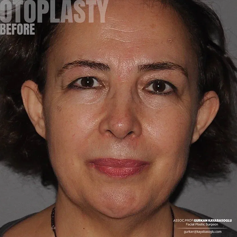 Before & after — Otoplasty (Ear Reshaping)
6. Otoplasty Recovery Dynamics & Safe Healing Timeline
Because the new ear shape is securely held by deep permanent internal stitches, the external skin heals quickly. The primary focus during recovery is protecting the ears from folding forward or experiencing accidental friction while the cartilage structure bonds in its new position.
Milestone Recovery Timeline
| Timeframe | Anatomical Healing Behavior | Clinical Care Priorities |
|---|---|---|
| Days 1 - 3 | The ears feel tight, tender, and noticeably swollen. Faint bruising may appear around the back folds. | Keep the firm clinic compression wrap completely dry. Take prescribed mild analgesics to manage initial throbbing. Avoid sleeping on your side. |
| Days 5 - 7 | Initial tissue throbbing subsides, and swelling begins to decrease. The incision behind the ear achieves secure surface bonding. | Visit the clinic to remove the bulky medical wrap. Switch to a soft, elastic sports headband to protect the ears. Gentle hair washing is permitted once cleared. |
| Weeks 2 - 3 | Most noticeable swelling and bruising fade away. The ears begin to look more natural and integrated into your facial frame. | Wear the protective headband continuously at night to prevent the ears from bending forward during sleep. You may return to light office work. |
| Weeks 4 - 6 | The internal stitches achieve deep, permanent structural stability. Cartilage tenderness disappears completely, and skin sensitivity returns to normal. | You can stop wearing the headband completely. Avoid contact sports, heavy lifting, or activities that risk pulling the ear framework. |
| Months 2 - 3 | Residual micro-swelling resolves entirely. The scar hidden behind the ear fades into a thin, pale line that is very difficult to see. | Enjoy the permanent, balanced position of your ears. The structural adjustments are now fully stable and long-lasting. |
Post-Operative Care Protocols for Safe Ear Contouring
Dos:
- Wear your protective elastic headband exactly as directed by your surgeon, especially during sleep, to shield the healing cartilage from folding.
- Keep the incision lines behind the ears completely dry and clean, applying only the specialized antibiotic ointments provided by the clinic.
- Sleep exclusively on your back with your head elevated on firm pillows to help reduce local swelling and prevent pressure on the ears.
Don’ts:
- Do not sleep directly on your side or rest your head on your ears for at least one month following the surgery.
- Do not pull, bend, or check the strength of your ear cartilage in the mirror, as this creates unnecessary tension on the fresh internal stitches.
- Do not expose your healing ears to direct heat sources like hair dryers, hot styling tools, or intense sunlight while skin sensitivity is temporarily reduced.
7. Cost & Logistics Analysis: Istanbul, Turkey
An otoplasty requires specialized surgical equipment and a fully equipped clinical facility to guarantee the highest level of patient safety. While the specific asymmetry and cartilage thickness shape your individual surgical layout, undergoing ear reshaping in Istanbul, Turkey, provides world-class facial plastic expertise combined with highly optimized, comprehensive pricing packages.
Factors Influencing Ear Reshaping Pricing
- Bilateral vs. Unilateral Correction: Reshaping both ears to fix overall protrusion requires different operating room time and surgical resources than correcting a single asymmetrical ear.
- Structural Combination Needs: Combining a standard ear fold lift with a conchal cartilage reduction to treat a very deep ear bowl alters the overall technical layout and shapes surgical fees.
- Anesthesia Protocol: Choosing between localized anesthesia with deep intravenous sedation or full general anesthesia inside an accredited surgical facility defines the base hospital facility charges.
Global Price Comparison Grid
| Country / Region | Estimated Average Cost Range (Bilateral) | Logistical Package Benefits |
|---|---|---|
| Istanbul, Turkey | €2,650 – €3,200 | All-inclusive coordination: Expert surgeon fees, accredited hospital facility costs, anesthesia, luxury hotel stay, and all private airport-clinic transfers. |
| United States (USA) | €9,300 – €11,200 | Surgeon fees are usually quoted alone; operating theater use, anesthesia supplies, and follow-up clinical visits frequently incur separate bills. |
| United Kingdom (UK) | €8,000 – €9,600 | High private medical facility overheads lead to substantial base rates for comprehensive ear adjustments. |
| Canada | €7,400 – €9,000 | Cosmetic facial surgery is outside public health coverage; surgeon, operating facility and anaesthesia are billed separately, and travel or accommodation is never part of the quote. |
| Australia | €8,500 – €10,200 | Medicare does not fund cosmetic indications; high theatre and anaesthetist charges sit on top of the surgeon’s quote, with no transfer or accommodation support. |
Travel and Healing Logistics for International Patients
Because ear reshaping involves fine skin sutures and requires balanced pressure management using a compression wrap during early recovery, ensuring initial tissue stability before flying home is essential. International patients should plan a stay of 6 to 7 days in Istanbul. This window allows the early ear swelling to resolve comfortably, enables direct evaluation of your initial healing, and ensures all fine external sutures behind the ears are safely removed or checked at the clinic before your flight back.
8. Prof. Dr. Gürkan Kayabaşoğlu’s Otoplasty Philosophy
A successful otoplasty should never result in an over-corrected, “plastered-to-the-head” appearance. Rejuvenating and reshaping the ears requires a deep respect for natural craniofacial proportions, ensuring the ears sit at a balanced angle while retaining their soft, organic curves when viewed from the front.
The Pillars of Our Ear Reshaping Approach
- Proportion Preservation Over Aggressive Flattening: Traditional methods often pulled the ears back as flat as possible, which erased the natural shadow folds and created an artificial, pinned-back look. Our approach focuses on custom angles, ensuring the outer rim remains beautifully visible and correctly spaced from the scalp.
- Maintaining Soft Cartilage Transitions: Rebuilding an ear fold should never create sharp, unnatural ridges on the outer surface. By utilizing advanced cartilage-sparing suture techniques instead of aggressive cutting, the new antihelical fold maintains a smooth, rounded contour that mimics natural genetics.
- Securing the Structural Foundation: Long-term stability relies on strong internal anchoring, not tight skin closure. We secure the structural cartilage layers directly to the stable bone frame behind the ear, absorbing the natural spring-back force of the tissue to prevent the ear from shifting forward later.
Otoplasty Surgical Application Matrix
| Structural Goal | Anatomical Challenge | Dr. Kayabaşoğlu’s Structural Solution |
|---|---|---|
| Natural Antihelical Curve | A flat, unfolded upper ear frame that causes the top rim to project outward. | Precise placement of permanent internal mattress sutures to guide the cartilage into a smooth, natural-looking fold. |
| Balanced Central Positioning | An overdeveloped, deep central ear bowl (conch) that pushes the middle ear frame away from the head. | Meticulous trimming of excess cartilage height combined with strong, permanent stitches anchored directly to the bone frame. |
| Symmetrical Ear Projection | Noticeable shape or angle differences between the left and right ears. | Customized adjustment vectors tailored for each ear during surgery to achieve a balanced, natural facial framework. |
9. Frequently Asked Questions (FAQ Matrix)
This comprehensive question-and-answer framework clarifies the anatomical, technical, and recovery details of advanced otoplasty procedures.
Will there be visible scars on my ears after an otoplasty?
No. The surgical incision is placed entirely within the natural post-auricular crease, which is the shadow fold located behind the ear where it meets the head. Because the incision follows this natural groove and is closed cleanly with fine sutures, the resulting scar remains completely hidden from both the front and side views.
Can the ears spring back or return to their old position after surgery?
This risk is minimized by using advanced deep-plane anchoring. If a surgeon relies only on skin tightening, the natural memory of the cartilage can cause the ear to drop back out. We prevent this by placing permanent, high-strength internal sutures directly into the cartilage folds and securing them to the firm bone lining behind the ear.
How long do I need to wear the headband after ear reshaping surgery?
Patients wear a firm compression dressing for the first 5 to 7 days until the initial clinic check. After this wrap is removed, you switch to a soft, elastic sports headband. This headband must be worn continuously during the second week, and then strictly at bedtime for an additional 2 to 3 weeks to prevent the ears from bending forward during sleep.
Is otoplasty suitable for young children, and what is the best age?
Yes, otoplasty is highly successful in children. The ideal age is between 5 and 6 years old. By this stage, the ear cartilage has reached roughly 85-90% of its adult size and remains highly flexible, making it easier to reshape. Correcting protrusion at this age also helps protect children from experiencing peer teasing before they enter primary school.
Is the procedure performed under local or general anesthesia?
For teenage and adult patients, an isolated otoplasty can be completed comfortably under local anesthesia combined with intravenous sedation, ensuring a completely pain-free and relaxed experience. For younger children, general anesthesia is preferred to guarantee that the child stays completely still and comfortable throughout the surgery.
When can I return to exercising, swimming, and playing contact sports?
You can resume light walking within a few days and return to light gym exercise or running after 3 weeks. However, swimming should be avoided for 4 weeks until the skin incisions behind the ears are fully sealed. Heavy contact sports like basketball, soccer, or martial arts must be avoided for at least 2 months to protect the structural stitches from trauma.